| |
Scientists often find it necessary to study the biological processes of proteins apart from the other reactions that occur in cells. In order to analyze proteins outside of a living plant, animal or microbe, they must first be purified away from other cell contents.
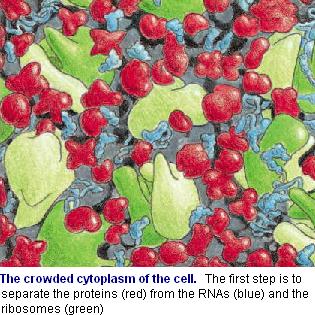
Generally speaking, the extraction of proteins from plant tissue begins by gently grinding the tissue and breaking the cell membranes so that their contents are released into a specific solution.
|
|
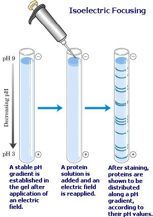
Scientists who study proteomics usually do this with a two-step technique called two dimensional gel electrophoresis.
The first step is isoelectric focusing. A special detergent is added to the solution of proteins to release the tangle of protein chains. These are then separated on the basis of their natural charge (their isoelectric point) by putting them in a narrow tube of polyacrylamide gel that contains a pH gradient. An electric current is applied down the length of the strip of gel. Over time, the different proteins move with the current but stop at different places in the gel.
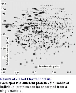
In the second step the proteins are separated on the basis of size, by using a technique called sodium dodecyl sulfate polyacrylamide-gel electrophoresis (SDS-PAGE). The narrow gel containing the separated proteins is again subjected to an electric current, but in a direction that is at a right angle to the direction that used in the first step. Each protein then migrates away from its fellows of different size to form a discrete spot. The gel is stained with a dye to make the spots of protein visible.
2-D gel electrophoresis can distinguish between two proteins that differ in only one charged amino acid.
|
|